1. A 40 year-old man presents to his physician with a 4 day history of terrible headache, fever, confusion, and staggering gait. His HIV status is unknown. A lumbar puncture is performed and CSF is sent to the laboratory. Which of the following results would you expect from this patient’s CSF studies:
a. Normal CSF pressure with a high protein level
b. India ink test with presence of encapsulated yeast
c. Elevated polymorphonuclear cells with high protein levels
d. Positive Galactomannan assay
e. Positive Ouchterlony-mannan test for Candida species
2. A yeast is identified in the laboratory with the following features; chlamydospore production, germ tube production and green pigment produced on ChromAgar. The yeast is identified as:
a. Candida dublinensis
b. Candida tropicalis
c. Candida glabrata
d. Candida albicans
e. Candida lusitaniae
3. A 32 year-old female presents to her physician with a hypo-pigmented skin lesion. A skin scraping was submitted to the laboratory for KOH preparation and fungal culture. The KOH examination was described as hyphae with yeast like structures in a spaghetti and meatball arrangement. The yeast did not grow on Sabouraud’s agar after 72 hours of incubation. This infection is most likely due to:
a. Candida albicans
b. Malassezia furfur
c. Trichophyton rubrum
d. Microsporum canis
e. Prototheca species
4. A 27 year-old man from Missouri presents to his physician with shortness of breath, fever and fatigue. In addition to his job as an accountant, he enjoyed spelunking and trout fishing thorough out the year. A sputum was submitted for fungal culture and a white mold grew after 14 days of incubation at 30*C. Under the microscope the mold appeared as septate thin hyphae with both microconidia and large tuberculate macroconidia. The mold is identified as:
a. Coccidioides immitis
b. Blastomyces dermatitidis
c. Histoplasma capsulatum
d. Fusarium species
e. Sporothrix schenckii
5. This organism was isolated from the sputum of a 45 year-old female bone marrow transplant patient. The photograph was taken from an organism grown on inhibitory mold agar at 30*C after 7 days of incubation. The identification is:

a. Fusarium species
b. Penicillium species
c. Acremonium species
d. Scopulariopsis species
e. Paecilomyces species
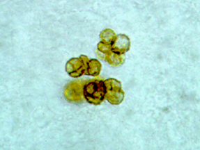
6. A 50 year-old male from Guatemala presented to his physician with a chronic skin lesion which overtime developed a cauliflower-like appearance. A biopsy was submitted for examination and revealed the structure seen in this photo. This structure is associated with what type of infection:
a. Phaeohyphomycoses
b. Chromoblastomycoses
c. Mycetoma
d. Sporotrichosis
e. Actinomycosis
7. A biopsy of an infected lung from a 50 year-old lung transplant patient revealed uniform hyphae with regularly spaced septation and branching at a 45 degree angle. No yeast cells were observed. Which of the following is the most probable diagnosis?
a. Actinomycosis
b. Aspergillosis
c. Blastomycosis
d. Cryptococcosis
e. Zygomycosis
8. A 56 year-old female complains of red nodular and ulcerative painful lesions on the right arm. She recalls being stuck with thorns while gardening. Her physician diagnosed a fungal infection. Which organisms is most likely the etiology?
a. Aspergillus fumigatus
b. Candida albicans
c. Nocardia asteroides
d. Sporothrix schenckii
e. Prototheca species
9. A 43 year-old female experienced fever and shortness of breath. She lived in the northwestern part of the US and spent time trekking through the forested areas of the region. Her sputum culture grew yeast on Sabouraud’s agar 8 – 10um in size, growth turned brown on birdseed agar, and blue on L-canavanine glycine brom-thymol blue medium.
a. Cryptococcus neoformans
b. Candida albicans
c. Cryptococcus gattii
d. Cryptococcus albidus
e. Candida dublinensis
10. A 45 year-old male complained of blockage of his nasal cavity by a slow growing lesion. He was a recent imigrant from Brazil. A biopsy of the lesion showed a large yeast cell with multiple buds appearing in a “mariners-wheel” type arrangement. Which organisms is the most likely etiology?
a. Blastomyces dermatitidis
b. Paracoccidioides brasileinsis
c. Prototheca species
d. Trichosporon beigelii
e. Cryptococcus albidus
Thank you very much for this informative and organized blog, it's a helpful tool for the board review. I couldn't find the key for mycology questions,are they posted somewhere else?
ReplyDeleteCheck with this link for this set with the answers: http://www.microbeswithmorgan.com/2015/11/mycology-questions-with-text-set1.html
ReplyDelete