Friday, December 4, 2015

Virology Review Questions

Virology Review Questions
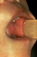
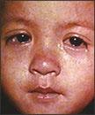
  1. A 10 year old boy was examined in the emergency room. He had a 24 hour history of fever (101.7*F), runny nose, sore throat, inflamed eyes and skin rash (large blotches).  Upon examination inside the mouth, tiny white spots with bluish-white centers were seen  This patient’s infection is most likely:

    1. Varicella –Zoster
    2. Rubella
    3. Rubeola
    4. Enterovirus
    5. Coronavirus
Koplik spots in measles. Photograph courtesy of WoMeasles conjunctivitis.

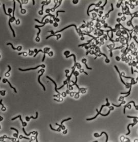
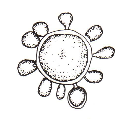
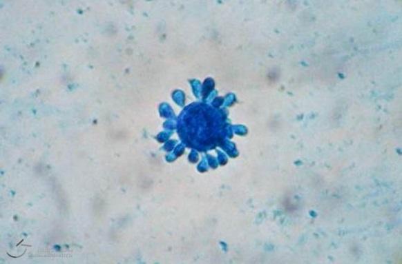
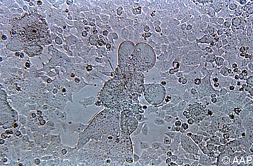
  1. A one year old boy visited his physician with coughing, sneezing, fever (102°F) and some difficulty breathing.  A nasal washing was collected and was sent to the laboratory for testing which included a viral cell culture.  The picture below is most consistent with which viral pathogen (Cytopathic effect in Hep-2 cell line):

    1. Rhinovirus
    2. Human metapneumovirus
    3. Coronavirus
    4. Respiratory Syncytial virus
    5. Parainfluenzae virus
http://o.quizlet.com/i/d_8IggGT7sjYqyf8_eltkA.jpg
  1. A 34 year old female became abruptly ill after arriving home from a three day cruise to Ensenada, Mexico.  She developed acute onset of vomiting and developed watery non-bloody diarrhea.  She heard that numerous travelers had also become ill following the cruise.  She started feeling better twenty four hours after symptoms began.  Her disease is most consistent with which pathogen:

    1. Norovirus
    2. Rotavirus
    3. Sapovirus
    4. Enterovirus
    5. Astrovirus

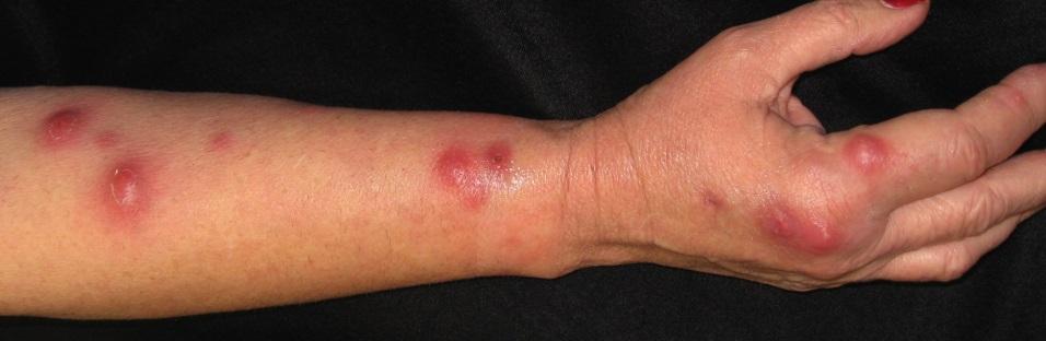
  1. A 40 year old male returned from the Caribbean one week prior.  He had a sudden onset of fever (102*F), intense joint pain, headache, and a maculo-papular rash.  While in the Caribbean he experirience many mosquito bites.  The best diagnostic test should include:

    1. Viral cell culture of nasal secretions
    2. IgG, IgM for Dengue virus (serum)
    3. Qualitative PCR for Chikungunya virus (blood)
    4. Quantitative PCR for Dengue virus (blood)
    5. Qualitative PCR for Chikungunya virus (urine)

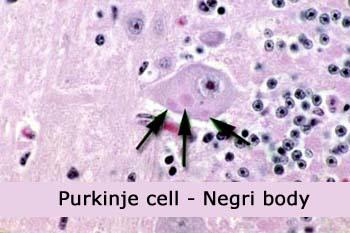
  1. A 16 year old girl returned from a trip to Mexico approximately one week prior and reported to her local Emergency Department (ED).  During her trip she encountered various animals along the way, including feral dogs and cats.  She approached a few of the animals and was involved in some skirmishes with small nips and scratches.  In the ED, she was found to have general weakness, slight fever and headache for the last few days.  Over the next week she progressed to a state of anxiety and confusion and a brain biopsy was performed.  This pathology is diagnostic for which infection:
    1. Cytomegalovirus    
    2. Rabies virus   
    3. Toxoplasma
      d.  Varicella Zoster
       e.  Herpes simplex

F21491.jpg (30324 bytes)


ANSWERS

  1. (c)  Measles is historically a childhood infection caused by the Rubeola virus. Once quite common, measles can now be prevented with a vaccine (MMR- Measles, Mumps and Rubella).  Measles can be serious and even fatal for small children. While death rates have been falling worldwide as more children receive the measles vaccine, the disease still kills more than 100,000 people a year, most under the age of 5.  Non-immunized adults are susceptible to infection and can experience complications.  Measles signs and symptoms appear 10 to 14 days after exposure to the virus. Signs and symptoms of measles include cough, runny nose, inflamed eyes (conjunctivitis), sore throat, fever and a red, blotchy skin rash. The skin rash is made of large, flat blotches that often flow into one another.  Also seen are tiny white spots with bluish-white centers on a red background found inside the mouth on the inner lining of the cheek — known as Koplik's spots. If laboratory diagnosis is necessary the tests of choice include serum IgM for measles antibody and qualitative PCR on a urine specimen.


  1. (d) Respiratory Syncytial Virus (RSV) causes infections of the lungs and respiratory tract. It's so common that most children have been infected with the virus by age 2. Respiratory syncytial virus can also infect adults.  In adults and older, healthy children, the symptoms of respiratory syncytial virus are mild and typically mimic the common cold.  Self-care measures are usually all that's needed to relieve any discomfort.  Infection with respiratory syncytial virus can be severe in some cases, especially in premature babies and infants with underlying health conditions. RSV can also become serious in older adults, adults with heart and lung diseases, or anyone with a very weak immune system (immunocompromised).  If laboratory testing is necessary, currently the most useful test is polymerase chain reaction using nasopharyngeal or nasal secretions (PCR).  In years past, viral cell culture was the method of choice.  The presence of RSV is confirmed by the formation of giant cells or syncytia formation in inoculated cell cultures in 1 – 2 weeks.

  1. (a)  Noroviruses are a group of viruses that cause inflammation of the stomach and large intestine lining (gastroenteritis); they are the leading cause of gastroenteritis in the U.S. The norovirus was originally called the Norwalk virus after the town of Norwalk, Ohio, the location of the first confirmed outbreak in 1972. People become infected with noroviruses when they eat food or drink liquids that have been contaminated. You can also become infected if you touch an object or surface that has been infected with the virus and then touch your nose, mouth, or eyes. Noroviruses thrive on cruise ships (as well as in day-care centers, restaurants, nursing homes, and other close quarters) because they are very hardy and highly contagious. They can survive temperature extremes in water and on surfaces.  Once someone is infected from contaminated food, the virus can quickly pass from person to person through shared food or utensils, by shaking hands or through other close contact. People who have a weakened immune system are particularly susceptible to catching noroviruses,  When infected with norovirus, you will be ill within a 24-48 hours with  nausea, vomiting (more often in children), watery diarrhea (more often in adults), and stomach cramps. If diagnostic testing is necessary, this virus cannot grow in cell culture and the method of choice is polymerase chain reaction (PCR) on stool or vomitus.

  1. (c)  Chikungunya is a viral disease that is transmitted by mosquitoes. It has occurred in Africa, Southern Europe, Southeast Asia, and islands in the Indian and Pacific Oceans,  In late 2013.  Chikungunya was found for the first time in the Americas on islands in the Caribbean.  Aedes species mosquitoes transmit chikungunya virus and this is the same genus of mosquitoes that transmit dengue virus.  These mosquitoes bite mostly during the daytime.  Symptoms usually begin 3‒7 days after being bitten by an infected mosquito and the most common symptoms are fever and severe joint pains, often in the hands and feet. Other symptoms may include headache, muscle pain, joint swelling, or rash.  Unlike Dengue, there are no hemorrhagic complications. There are no antiviral medicines to treat chikungunya.  Most patients improve after 7 days. People at increased risk for severe disease include newborns exposed during delivery, older adults (≥65 years), and people with medical conditions such as high blood pressure, diabetes, or heart disease, but deaths are rare.

  1. (b)  Rabies virus belongs to the family Rhabdovirid and have a distinct "bullet" shape that can be seen on EM.  The first symptoms of rabies may be very similar to those of the flu including general weakness or discomfort, fever, or headache. These symptoms may last for several days.  There may be also discomfort or a prickling or itching sensation at the site of bite, progressing within days to symptoms of cerebral dysfunction, anxiety, confusion, agitation. As the disease progresses, the person may experience delirium, abnormal behavior, hallucinations, and insomnia. The acute period of disease typically ends after 2 to 10 days. Once clinical signs of rabies appear, the disease is nearly always fatal, and treatment is typically supportive. Disease prevention includes administration of both passive antibody, through an injection of human immune globulin and a round of injections with rabies vaccine.  Histopathologic evidence of rabies encephalomyelitis (inflammation) in brain tissue and meninges includes the following:  Mononuclear infiltration, Perivascular cuffing of lymphocytes or polymorphonuclear cells, Lymphocytic foci, and Negri bodies.

References:  
Centers for Disease Control and Prevention, CDC Home site.
Manual of Clinical Microbiology, 10th Edition, 2011.  Editors: James Versalovic1, Karen C. Carroll2, Guido Funke3, James H. Jorgensen4, Marie Louise Landry5, David W. Warnock

Thursday, November 19, 2015

Mycology Questions Set #2 with Text

Mycology Questions Set #2 with Text ANSWERS
6. A 70 yo male presents to a clinic visit with shortness of breath and fatigue.  He is 6 months status post left lung transplant.  A CT scan shows consolidation in the left lower lobe in addition to nodules throughout the left lobe.  Serial serum 1-3-Beta-D-glucan and Aspergillus galactomannan testing is negative.  Bronchcoscopy is ordered with biopsy.  Histological examination of the biopsy shows the following on Gomori Methanamine stain (GMS).  What is the most likely fungal organism in the tissue:
https://bioweb.uwlax.edu/bio203/s2012/lind_vale/WEBSITE%20PHOTOS/Mucor%20zygomytosis.jpg
a. Spergillus
b.  Scedosporium   
c.  Mucor
d.  Candida
f.  Fusarium
7.  A 27 year-old man from Missouri presents to his physician with shortness of breath, fever and fatigue.  In addition to his job as an accountant, he enjoys spelunking and trout fishing. A sputum was submitted for fungal culture and a white mold grew after 14 days of incubation at 30*C.  An adhesive tape prep revealed thin septate hyphae with both microconidia and large tuberculate macroconidia.  The mold is identified as:
rounded tuberculate macroconidia
a. Coccidioides immitis
b. Blastomyces dermatitidis
c. Histoplasma capsulatum
d. Fusarium species
e. Sporothrix schenckii


8.  A 43 year-old female experienced fever and shortness of breath.  She lived in the northwestern part of the US and spent time trekking through the forested areas of the region.  Her sputum culture grew a mucoid yeast colony on Sabouraud’s agar, the yeast was 8 – 10um in size, growth turned brown on birdseed agar, and blue on L-canavanine glycine brom-thymol blue medium.  This yeast can be identified as:
R01192
a. Cryptococcus neoformans
b. Candida albicans
c. Cryptococcus gattii
d. Cryptococcus albidus
e. Candida dublinensis


9. A 32 year-old female presents to her physician with a hypo-pigmented skin lesion.  A skin scraping was submitted to the laboratory for KOH preparation and fungal culture.  The KOH examination was described as hyphae with yeast like structures in a spaghetti and meatball arrangement.  The yeast did not grow on Sabouraud’s agar after 72 hours of incubation but grew after the addition of oil to the culture plate.  This infection is most likely due to:
10% KOH with Parker ink mount

a. Candida albicans 

b. Malassezia furfur

c. Trichophyton rubrum

d. Microsporum canis

e. Prototheca species

10.  A yeast was isolated on Sabourauds agar after 24 hrs incubation at 30°C. The yeast produced chlamydospores on cornmeal agar, germ tubes were produced with incubation in serum at 35°C for 4 hours, and green pigmentation was produced on chromogenic agar.  These reactions identify the yeast:


http://www.pfdb.net/photo/makimura_k/box20010915/standard/i_04z.jpg http://www.pfdb.net/photo/iwaguchi_s/box1/standard/germ_tube-1.jpg http://www.pfdb.net/photo/makimura_k/CHROMagar/standard/C_albicans_ATCC10231.jpg
a. Candida dublinensis
b. Candida tropicalis
c. Candida glabrata
d. Candida albicans
e. Candida lusitaniae


ANSWERS

6. (c) Mucor.  This patient has an invasive infection with Mucor species.  Mucor is a member of the of the rapidly growing fungi known as the Zygomycetes  (most commonly Mucor, Rhizopus, Absidia, Cunnignhamella species) that are ubiquitous in the environment. These fungi grow rapidly on most fungal media producing aerial mycelia. They can produce have a wide range of clinical manifestations in humans, from cutaneous infection to sinusitis to pneumonia. The presence of a black necrotic eschar either in the nasopharynx or palate is a useful clue for this type of fungal infection.  In disseminated infections the prognosis is very poor as these are rapidly growing and incredibly invasive pathogens.  Major risk factors include diabetes and receipt of solid organ or stem cell transplant.  In transplant recipients, incidence occurs late in the post-transplant stage (usually greater than 3 months), as was the case for this patient
Based on the appearance on GMS and lack of positivity on serial 1-3-Beta-D-glucan testing, the only possible correct choice for part one was Mucormycosis.  The other infections listed should be positive for 1-3-Beta-D-glucan.  Further, the appearance of wide, ribbon-like hyphal elements on GMS is also characteristic of Mucormycosis.  This GMS appearance would also be consistent with the other member of the Zygomycetes, which include Rhizopus and Absidia species.
7. (c) Histoplasma capsulatum exhibits thermal dimorphism by growing in tissue or in culture at 37C as a budding yeast or in soil or culture at temperatures below 30C as a mold. On Sabouraud's agar at 30°C, colonies are slow growing, white or buff-brown, suede-like to cottony. Microscopic morphology shows the presence of characteristic large (8-14 um in diameter), rounded, single-celled, tuberculate macroconidia formed on hyaline hyphae. Microconidia, if present, are small (2-4 um in diameter), round and on short branches of the hyphae.  On brain heart infusion (BHI) blood agar incubated at 37°C, colonies are smooth, moist, white and yeast-like. Microscopically, numerous small round to oval budding yeast cells, 3-4 x 2-3 um in size are observed.
The fungus lives in the environment, particularly in soil that contains large amounts of bird or bat droppings. In the United States, Histoplasma mainly lives in the central and eastern states, especially areas around the Ohio and Mississippi River valleys. The fungus also lives in parts of Central and South America, Africa, Asia, and Australia. Infection occurs from breathing in the microscopic fungal spores from the air. Although most who breathe in the spores don’t become infected (95%), those who do may develop a fever, cough, and fatigue. Many infected recover on their own without medication, but those with weakened immune systems can develop pulmonary infection or disseminate to organs of the reticuloendothelial system.
8. (c) Cryptococcus gattii is a yeast that lives in the environment in many tropical and sub-tropical areas of the world as well as British Columbia and the U.S. Pacific Northwest. Cases have also been reported in California. C. gattii cryptococcosis is a rare infection, occurong from breathing in the yeast cell from nature.  The natural reservoir of C. gattii has yet to be fully revealed but some evidence points to forested areas and trees and possibility. It is not associated with pigeon excreta like C. neoformans.  The infection can affect both the lungs, where it can produce a cryptococcoma, and the central nervous system.  Both C. neoformans and C. gattii turn brown with growth on bird seed agar but only C. gattii will turn blue on L-canavanine glycine brom-thymol blue medium.
http://www.mycology.adelaide.edu.au/images/crypto5.gifhttp://www.mycology.adelaide.edu.au/images/crypto1.gif


9. (b) Malassezia furfur is the causative agent of Pityriasis versicolor, Pityriasis folliculitis and it has recently been implicated as a causative agent of seborrhoeic dermatitis and dandruff. It has also been recovered in blood cultures from neonate and adult patients undergoing lipid replacement therapy. M. furfur is a lipophilic yeast. Pityriasis versicolor is a chronic, superficial fungal disease of the skin characterized by white, pink, or brownish lesions,and covered with thin furfuraceous scales. Lesions occur on the trunk, shoulders and arms, rarely on the neck and face, and fluorescent pale greenish color under Wood's ultra-violet light. Young adults are affected most often, but the disease may occur in childhood and old age.
Skin scrapings taken from patients with Pityriasis versicolor when mounted in 10% KOH show characteristic clusters of thick-walled round, budding yeast-like cells with collarettes and short angular hyphal forms. These microscopic features are diagnostic for Malassezia furfur and culture preparations are usually not necessary.  If culture is performed, oil must be added to the culture media to promote growth.
http://www.parmoon.co/en/wp-content/uploads/2013/05/untitled.jpghttp://howmed.net/wp-content/uploads/2013/08/Scanning-electron-micrograph-of-Malassezia-furfur.jpg
GMS stained skin biopsy showing characteristic spherical yeast cells and short pseudohyphal elements typical of M. furfur that have been described as spaghetti and meatballs.
10. (d) Candida albicans is one of nine species of Candida which most commonly cause human infections.  It can be found in soil, inanimate objects, and foods.  It is also found as normal flora of the human GI tract, vagina, and skin and it is considered an opportunistic human pathogen.  
C. albicans grow well on many different agars and colonies are smooth, creamy white colonies.  Identification of C. albicans can be made by the observation of germ tubes, produced when C. albicans grows in serum.  C. albicans can also be differentiated from other yeast based on the microscopic morphology on corn meal agar.  C. albicans produce chlamydospores, large round structures produced along the pseudohyphae.  C. albicans produces a green pigment when grown on ChromAgar for Candida species.
http://upload.wikimedia.org/wikipedia/commons/thumb/f/f5/C_albicans_en.jpg/230px-C_albicans_en.jpg C-albicans Germ tube LAb11D-6b 2012

Mycology Questions with Text Set1


Mycology Questions with Text (Set 1)

1. This organism was isolated from the sputum of a 45 year old female bone marrow transplant patient.  The photograph is an adhesive tape preparation of a mold growing on inhibitory mold agar at 30°C after 7 days of incubation.  The identification is:

a. Fusarium species
b. Penicillium species
c. Acremonium species
d. Scopulariopsis species
e. Paecilomyces species

2. A 50 year-old male from Guatemala presented to his physician with a chronic skin lesion which overtime developed a cauliflower-like appearance.  A biopsy was submitted for examination and revealed the structure seen in this photo.  This structure is associated with what type of infection:


a. Phaeohyphomycoses

b. Chromoblastomycoses

c. Mycetoma

d. Sporotrichosis

e. Actinomycosis


  1. A lung biopsy from a 50 year-old lung transplant patient revealed uniform hyaline hyphae with regularly spaced septations and branching at a 45 degree angle.  No yeast cells were observed.  Which of the following is the most probable diagnosis?

    a. Actinomycosis

    b.Aspergillosis

    c. Blastomycosis

    d. Cryptococcosis

    e. Zygomycosis


4. A 56 year-old female visited her physician because of red nodular and ulcerative painful lesions on her right arm.  She recalls being stuck with rose thorns while gardening.  A yeast-like colony grew in culture after 5 days of incubation at 30°C.  The colony progressively turned black over 7 days.  Which organism is the most likely etiology?

a. Aspergillus fumigatus

b. Candida albicans

c. Nocardia asteroides

d. Sporothrix schenckii

e.Prototheca species


5.  A 45 year-old male complained of a slow growing lesion in his nasal cavity.  He was a recent immigrant from Brazil.   A biopsy of the lesion stained with Gomori Methenamine Stain (GMS) showed a large yeast cell (20 um) with multiple small buds appearing in a “mariners-wheel” type arrangement.  Which organisms is the most likely etiology?
a. Blastomyces dermatitidis
b. Paracoccidioides brasiliensis
c. Prototheca species
d. Trichosporon beigelii
e. Cryptococcus albidus

ANSWERS

1.    (a) Fusarium is a filamentous fungus widely distributed on plants and in the soil as well as being a common contaminant and a well-known plant pathogen.  The genus Fusarium currently contains over 20 species. The most common of these are Fusarium solani, Fusarium oxysporum, and Fusarium chlamydosporum   Fusarium is one of the emerging causes of opportunistic mycoses.  Fusarium spp. are causative agents of superficial and systemic infections in humans. The most virulent Fusarium spp. is Fusarium solani.  Trauma is the major predisposing factor for development of cutaneous infections due to Fusarium strains. Disseminated opportunistic infections, on the other hand, develop in immunosuppressed hosts, particularly in neutropenic and transplant patient. Fusarium infections following solid organ transplantation tend to remain local and have a better outcome compared to those that develop in patients with hematological malignancies and bone marrow transplantation patients.

  1. (b)  Chromoblastomycosis is a mycotic infection of the cutaneous and subcutaneous

    Tissues characterized by the development in tissue of dematiaceous (brown-pigmented), planate-dividing, rounded sclerotic bodies. Infections are caused by the traumatic implantation of fungal elements into the skin and are chronic, slowly progressive and localized.  Tissue proliferation usually occurs around the area of inoculation producing crusted, verrucose, wart-like lesions. World-wide distribution but more common in bare footed populations living in tropical regions.

    Etiological agents include various dematiaceous fungi associated with decaying vegetation or soil, especially Phialophora verrucosa, Fonsecaea pedrosoi, F. compacta and Cladophialophora carrionii Lesions of chromoblastomycosis are most often found on exposed parts of the body and usually start as a small scaly papules or nodules which are painless but may be itchy and as the disease develops rash-like areas enlarge and become raised irregular plaques that are often scaly or verrucose. In long standing infections, lesions may become tumorous and even cauliflower-like in appearance

https://blogger.googleusercontent.com/img/b/R29vZ2xl/AVvXsEi3ATTJUxeCIWy3x8J64TFnCCapIqoRiAAG_I07e895gqbnrSS2VjDHfm0aMgfccR1p3GVRDt6SkSknd8IllBgLfnvW6x-GOu__wE6QuOipUGKZnAPm-zqPL4ndD08sunZWzbz_s2BVfZs_/s400/untitled.bmp

  1. (b)  Aspergillus.  

    Although most people are often exposed to Aspergillus, infections caused by the fungus rarely occur in people who have a healthy immune system. The fungus is commonly found growing on dead leaves, stored grain, compost piles, or in other decaying vegetation. 

    There are several forms of aspergillosis:

    Allergic bronchopulmonary type that is an allergic reaction to the fungus. This infection usually develops in people who already have lung problems such as asthma or cystic fibrosis.

    Aspergilloma is a growth (fungus ball) that develops in an area of past lung disease or lung scarring such as tuberculosis or lung abscess.

    Pulmonary aspergillosis is an invasive type that is a serious infection with pneumonia that can spread to other parts of the body. This infection almost always occurs in people with a weakened immune system due to cancer, AIDS, leukemia, an organ transplant, chemotherapy, or other conditions or medications that lower the number or function of normal white blood cells or weaken the immune system.

    The morphology of Aspergillus  is better appreciated on special stains such as GMS and shows thin, septate hyphae with regular septations. The angle of the regular branching is at 45 degrees.
http://www.stritch.luc.edu/lumen/MedEd/mech/sgcases/case19/7a.jpg

  1. (d)  Sporothrix schenkii.  In nature Sporothrix lives as a saprophyte on wood, decaying vegetation (including rose thorns), Sphagnum moss, animal excreta and soil. The characteristic infection with Sporothrix involves suppurating subcutaneous nodules that progress along lymphatic channels (lymphocutaneous sporotrichosis). Primary pulmonary infection can occur (although rare) from inhalation of fungal spores (pulmonary sporotrichosis). In rare cases, disseminated S schenckii infection (disseminated sporotrichosis) occurs, characterized by disseminated cutaneous lesions and involvement of multiple visceral organs; this occurs most commonly in persons with AIDS.
http://escholarship.org/uc/item/4zx6v9j3/1.jpg
  1. (b)  Paracoccidioides brasiliensis
Paracoccidioidomycosis is a chronic granulomatous disease that characteristically produces a primary pulmonary infection, often inapparent, and then disseminates to form ulcerative granulomata of the buccal, nasal and occasionally the gastrointestinal mucosa.  The only etiological agent, P. brasiliensis is found in South and Central America. Microscopically, numerous large, 20-60 um, round, narrow base budding yeast cells are present. Single and multiple budding occurs, the latter are thick-walled cells that form the classical "steering wheel" or "Mariners wheel” morphology.


https://classconnection.s3.amazonaws.com/805/flashcards/151805/jpg/microscopic_morphology_of_paracoccidioides_brasiliensis_at_37c1332804383551.jpghttps://www2.ufmg.br/var/storage/imagensdoconhecimento/images/imagens/areas/ciencias-biologicas/enzima-patricia-campi/patricia-campi-imagem-5/8310-1-por-BR/Patricia-Campi-Imagem-5.jpg


Reference:
Versalovic, J, et al. Fungi, In: Manual of Clinical Microbiology, 10th ed., ASM Press; 2011.